

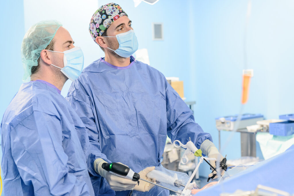
Minimally invasive surgery allows hernias to be repaired and the abdominal wall to be reconstructed through small incisions. Laparoscopy, the eTEP and PeTEP endoscopic techniques, and robotic assistance offer different ways of reaching the defect and positioning the surgical mesh. The aim is to restore the function of the abdominal wall and improve the pain or limitations caused by the hernia.
The surgical technique is chosen individually, taking into account the hernia, your health and your previous medical and surgical history. During your consultation, we agree on a treatment plan and explain its benefits, risks and recovery.
On this page: Types of hernia · Minimally invasive techniques · Recovery · Request an assessment
Inguinal and incisional hernias: two different problems
Inguinal and femoral hernias
These develop in the groin, where there are natural passageways through the abdominal wall. The lump may become larger when you stand, cough or lift something heavy, and smaller when you lie down. An examination distinguishes an inguinal hernia from a femoral hernia and checks whether one or both groins are affected. This distinction matters: femoral hernias carry a higher risk of complications and usually require a planned repair without a prolonged wait.
Read about the diagnosis and treatment of inguinal hernias.
Incisional hernias
An incisional hernia develops in the scar from a previous operation, including the scar from a small laparoscopic incision. Unlike an inguinal hernia, planning needs to take into account the condition of the abdominal wall after that surgery: the width of the defect, muscle quality, internal scar tissue and any previously implanted mesh. Repair may require reconstruction of a large part of the abdominal wall, extending beyond the visible lump.
Read about endoscopic and robotic reconstruction for incisional hernias.
Umbilical and epigastric hernias
An umbilical hernia develops at the navel; an epigastric hernia develops in the midline between the navel and the breastbone. Several small openings may be present, or there may also be rectus diastasis, a separation between the central abdominal muscles. Distinguishing these conditions helps us plan which area needs repair and how extensive the reinforcement should be.
Read about umbilical hernias, epigastric hernias and rectus diastasis.
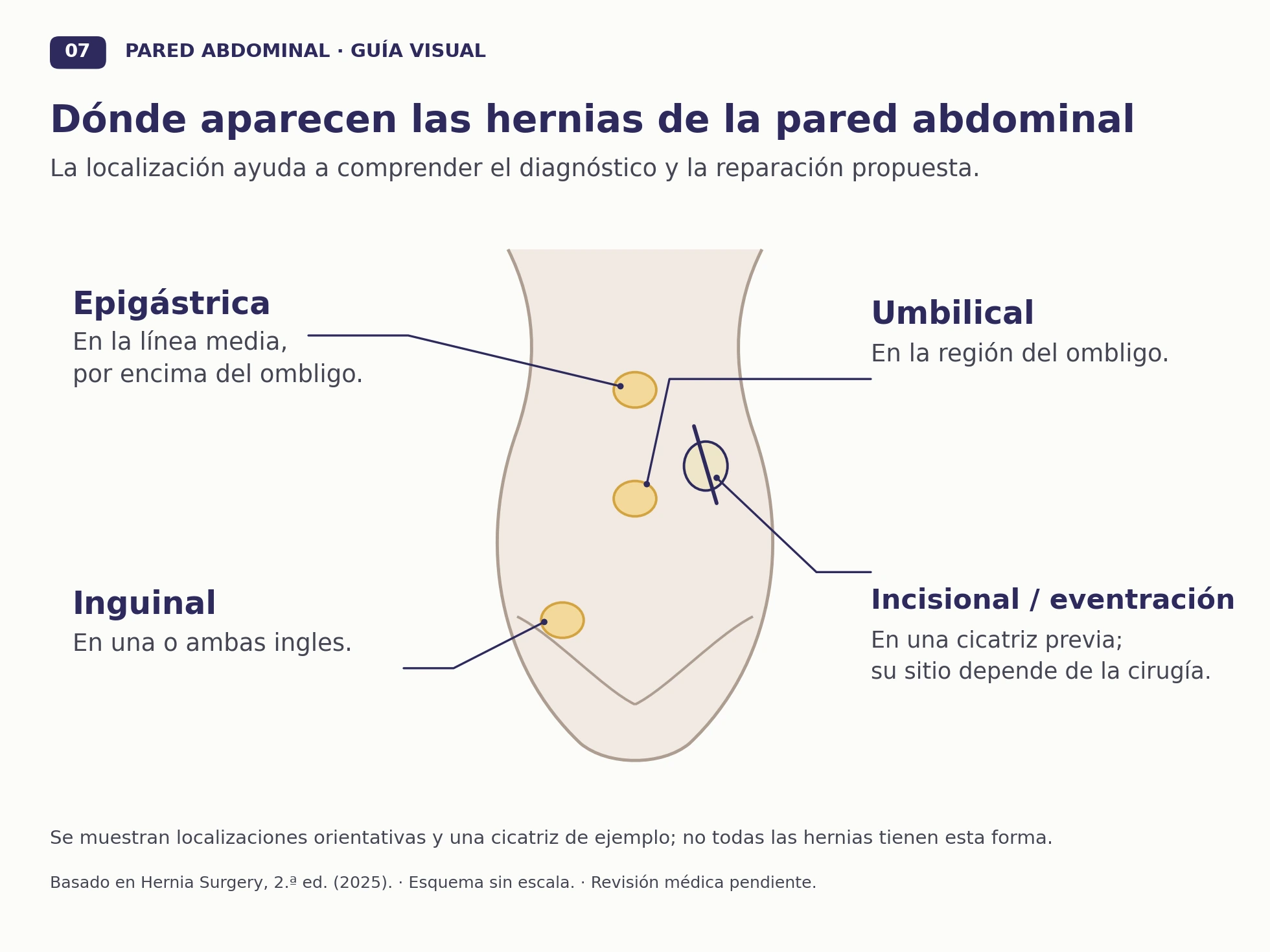
Enlarge the hernia location diagram.
What happens during a repair?
The operation returns the protruding tissue to the abdomen, repairs the defect in the abdominal wall and reinforces the weakened area. Many repairs use surgical mesh: an implant that becomes incorporated into the tissues and distributes strain over an area larger than the original opening. Mesh is not a surgical technique in itself; closing the defect, positioning the mesh and choosing how to reach the hernia also matter.
Laparoscopy, endoscopic reconstruction and robotic surgery
Reconstruction through small incisions
Laparoscopy and endoscopy use a camera and fine instruments. In the groin, TAPP reaches the repair space from inside the abdomen, whereas TEP approaches it from outside the peritoneal cavity. For incisional hernias, eTEP Rives–Stoppa creates a space behind the rectus muscles, while PeTEP works in the preperitoneal plane, in front of the parietal peritoneum, the membrane lining the inner surface of the abdominal wall. Both aim to position the mesh outside the peritoneal cavity.
Robotic assistance
The surgeon controls a camera and articulated instruments from a console. This platform helps with certain dissection and suturing movements, but it does not operate independently. It can be used for repairs such as inguinal rTAPP and eTEP or PeTEP approaches to the abdominal wall when the equipment is available. The name of the approach describes the repair; the robot is the tool used to carry it out.
Open surgery
Open surgery reaches the hernia through an incision over the area to be repaired. It includes the Lichtenstein technique for groin hernias and preperitoneal or retromuscular repairs for ventral and incisional hernias. In extensive reconstructions, it allows direct access to the scar and the different layers of the abdominal wall.
What we assess before surgery
Assessment starts with an examination while you are standing and lying down, a review of your symptoms and a discussion of previous operations. Ultrasound can help when the diagnosis is uncertain; for incisional hernias, a CT scan provides a map of the abdominal wall to measure the defect and plan reconstruction. We also review smoking, diabetes, weight and nutritional status, as these affect healing.
Bring reports from previous operations, any available scans and a list of your medicines. Explain which activities your hernia limits and whether your work involves physical effort: this helps us define the goals of treatment and plan your return to activity.
Recovery and follow-up
Walking, controlling pain and caring for the wound are part of recovery from the first few days. Returning to a desk job and returning to heavy lifting are different goals. Before discharge, you will receive advice about activity and follow-up appointments. The extent of the reconstruction, rather than the size of the skin incisions, helps explain how long recovery may take.
Risks include a haematoma, a seroma, infection, persistent pain and recurrence of the hernia. Follow-up allows us to assess wound healing and identify problems that need treatment.
When to seek emergency care
A lump that becomes very painful, no longer goes back in, or is accompanied by vomiting, abdominal swelling or changes in skin colour needs urgent medical attention. Do not wait for an online reply.
Abdominal wall consultation in Valencia
Dr Pablo Aragó Chofre is a specialist in General and Digestive Surgery and practises at IMED Valencia. A consultation allows us to confirm the diagnosis and explain the problem, the proposed repair and the next steps. Read about his professional background and training.
Request an appointment at your preferred IMED centre: Valencia (Burjassot), Colón or Gandía. In the booking portal, select Cirugía General y del Aparato Digestivo (General and Digestive Surgery) and Dr Pablo Aragó Chofre. If you need help or cannot find his appointments, see the telephone numbers and locations of the three centres.
The button opens IMED's external booking service, which has its own terms and privacy policy. Your appointment is subject to confirmation by the centre. Do not use this channel for emergencies.
More about abdominal wall surgery: Inguinal hernia: TEP, TAPP and rTAPP · Incisional hernia: endoscopic and robotic reconstruction · Umbilical and epigastric hernias
Medical sources
HerniaSurge: groin hernia guidelines, 2023 update and EHS: midline incisional hernia guidelines, 2023.
Review of the evidence on PeTEP, 2026.
Anatomical basis of the diagrams: Novitsky YW (ed.), Hernia Surgery: Current Principles, 2nd edition, 2025. Original illustrations based on the book; they do not reproduce its figures.