

Abdominal wall surgery: see all conditions and techniques
Medical information: Dr Pablo Aragó Chofre, specialist in General and Digestive Surgery. Read about his professional background.
An umbilical or epigastric hernia may be a small, isolated opening or part of a wider weakness of the abdominal midline. The PeTEP and REPA endoscopic techniques allow the hernia and any associated rectus diastasis to be repaired through small incisions. They work in different layers of the abdominal wall and place the reinforcement outside the peritoneal cavity. Planning the operation properly requires an assessment of the defect, not just the lump.
The surgical technique is chosen individually, taking into account the hernia, your health and your previous medical and surgical history. During your consultation, we agree on a treatment plan and explain its benefits, risks and recovery.
On this page: Hernias and rectus diastasis · PeTEP and REPA · Recovery · Request an assessment
Umbilical hernia, epigastric hernia and rectus diastasis: what is the difference?
Where the hernia develops
An umbilical hernia develops at the navel; an epigastric hernia develops in the midline between the navel and the breastbone. In both cases, there is a defect through which tissue, often fat, protrudes. It may cause discomfort when you strain, cough or press on the area. A small, painful lump does not always mean there is a large defect.
Rectus diastasis
In rectus diastasis, the gap between the rectus muscles of the abdomen becomes wider. This may form a central ridge when you sit up, but it is not itself a hernia opening. It can occur alongside an umbilical or epigastric hernia. When both are present, the whole midline needs to be assessed to decide how extensive the repair should be.
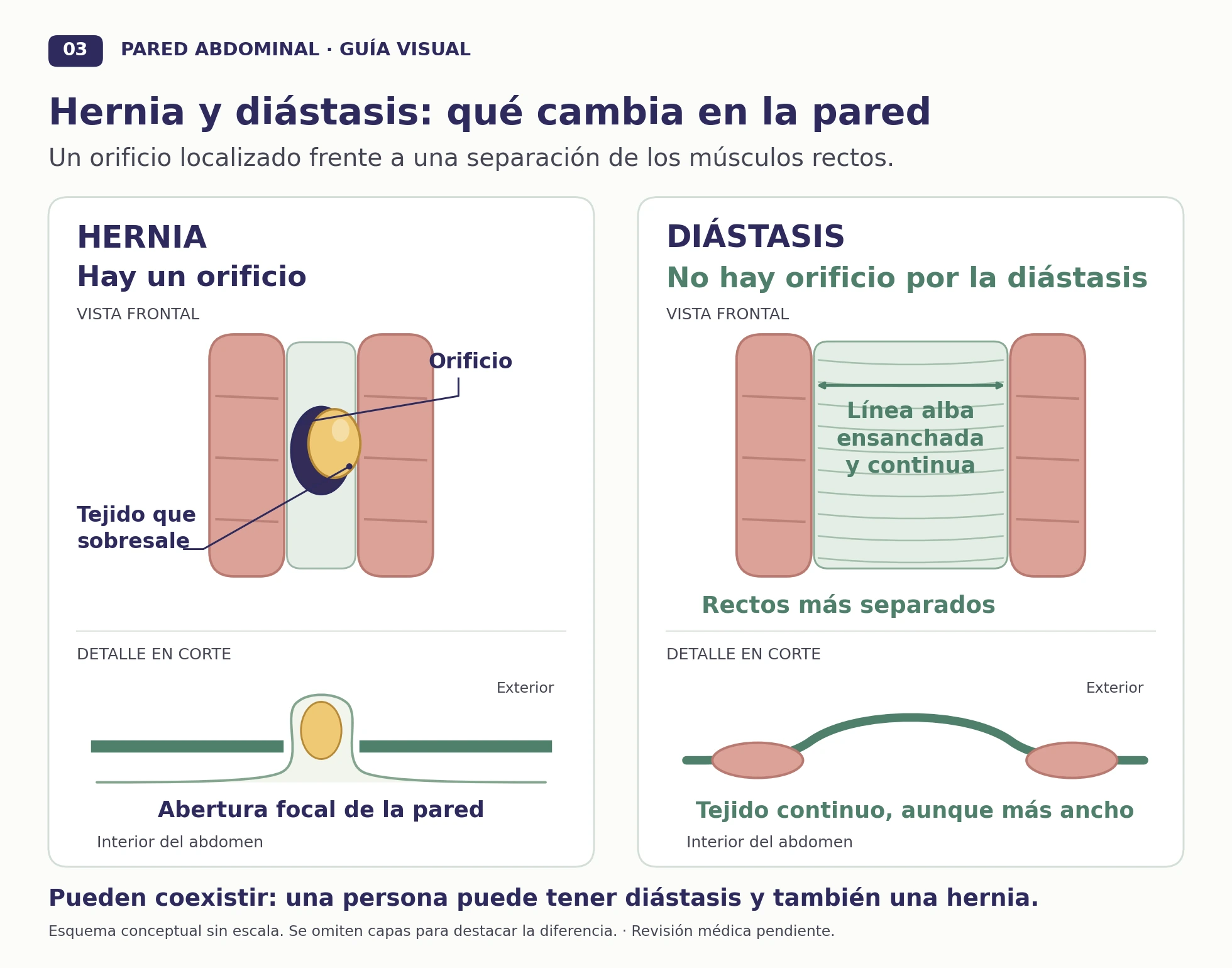
Enlarge the comparison of a hernia and rectus diastasis.
What is checked during the consultation?
The examination identifies the size and location of the defect, whether its contents go back in and whether there are other nearby openings. We also check for rectus diastasis and review previous operations. Ultrasound can help when the diagnosis is uncertain; more extensive imaging may be needed to plan a large reconstruction.
Pain, growth of the hernia and limitations in daily life help guide the decision about surgery. A small hernia that causes no discomfort can be monitored with follow-up. In adults, the defect does not usually close by itself: if the lump disappears when you lie down, this means its contents have gone back in, not that the hernia has healed.
PeTEP and REPA: two endoscopic ways to repair the midline
When an umbilical or epigastric hernia occurs with rectus diastasis, surgery can address both the hernia opening and the widening of the linea alba. PeTEP and REPA allow this to be done through small incisions, but they approach different layers of the abdominal wall.
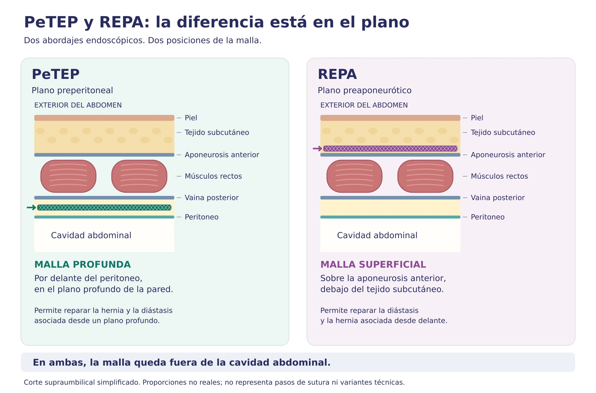
Enlarge the PeTEP and REPA diagram.
PeTEP: repair in the preperitoneal plane
PeTEP uses a totally extraperitoneal route: the working space is created within the abdominal wall, without using the cavity containing the abdominal organs as the usual route of access. The defect is closed and, when necessary, the midline is reconstructed with stitches. The mesh lies in a deep plane, in front of the peritoneum — the membrane lining the abdominal cavity — and separate from the bowel.
It is an option for small or medium-sized midline hernias, with or without rectus diastasis, when the aim is to reinforce the repair from a deep plane while preserving the space behind the rectus muscles. Whether this plane can be developed depends on the condition of the peritoneum, scars and mesh from previous operations. It is also used for some hernias that develop in surgical scars, known as incisional hernias.
REPA: endoscopic preaponeurotic repair
REPA works beneath the skin and in front of the aponeurosis, the fibrous layer covering the abdominal muscles. Through small incisions in the lower abdomen, stitches bring the widened midline together — a step known as plication — and the associated hernia is repaired. Mesh reinforcement is placed over this fibrous layer, in a more superficial position than in PeTEP.
Its main use is for rectus diastasis accompanied by an umbilical or epigastric hernia, especially when there is no significant excess skin that needs to be removed. It allows both problems to be treated in the same operation, but it does not remove excess skin and is not the same as an abdominoplasty.
Separating the tissue beneath the skin creates a space in which fluid can collect, forming a seroma. For this reason, drainage and care of this area are part of postoperative planning.
What matters when choosing between them?
The main difference is the repair plane and mesh position: deep and preperitoneal in PeTEP; superficial and preaponeurotic in REPA. Both can treat a hernia associated with rectus diastasis. For a small, isolated hernia without diastasis, a large area of the midline does not always need to be reconstructed.
There is not enough evidence to state that either technique is better for every patient. Long-term follow-up data for PeTEP are still limited; in REPA, seroma is a particularly relevant complication.
Why mesh is used
Closing an opening with stitches alone concentrates force at its edges. Mesh reinforces a wider area and reduces the risk of recurrence in many adult repairs. The size of the defect and the quality of the midline help determine how much tissue needs reinforcement. Some very small defects can be repaired with stitches alone.
The position of the implant also matters. In PeTEP it lies in front of the peritoneum; in REPA it lies over the anterior aponeurosis of the rectus muscles; and in retrorectus repairs it lies behind those muscles. These are different anatomical planes.
Open repair still has a role
For a small, localised hernia, an incision close to the navel may allow a direct repair without creating a large working space. Open preperitoneal repair is an established technique: it repairs the defect and places a flat mesh in front of the peritoneum. A retromuscular or sublay repair provides another plane of reinforcement behind the rectus muscles.
Preparation and returning to activity
Before surgery, your medicines, smoking, diabetes control and other factors that affect healing are reviewed. If you have had a previous operation or have mesh in place, bring the report: it helps identify which tissue planes have already been used.
Recovery includes walking, controlling pain and caring for the wound. Returning to a desk job and returning to sport or heavy lifting are planned separately. If a large area of the midline is reconstructed, small external incisions do not mean that the internal repair has already healed.
Swelling, a haematoma or a seroma may develop. A lump after surgery is not always a new hernia and should be assessed before drawing conclusions. There are also risks of infection, persistent pain and recurrence.
Warning signs
Seek urgent medical attention if the hernia becomes very painful, no longer goes back in, or is accompanied by vomiting, abdominal swelling or changes in skin colour. After surgery, seek medical advice for fever, wound discharge or pain that is getting worse rather than improving.
Assessment of an umbilical or epigastric hernia in Valencia
The consultation helps distinguish an isolated hernia from a wider problem affecting the midline and explain how each component would be repaired. Read about Dr Pablo Aragó's professional background.
Request an appointment at your preferred IMED centre: Valencia (Burjassot), Colón or Gandía. In the booking portal, select Cirugía General y del Aparato Digestivo (General and Digestive Surgery) and Dr Pablo Aragó Chofre. If you need help or cannot find his appointments, see the telephone numbers and locations of the three centres.
The button opens IMED's external booking service, which has its own terms and privacy policy. Your appointment is subject to confirmation by the centre. Do not use this channel for emergencies.
More about abdominal wall surgery: Abdominal wall surgery · Incisional hernia: a hernia in a surgical scar
Medical sources
EHS/AHS guidelines for umbilical and epigastric hernias, 2020. This information provides general guidance and does not replace an individual assessment.
Review of the evidence on PeTEP, 2026.
Anatomical basis of the diagrams: Novitsky YW (ed.), Hernia Surgery: Current Principles, 2nd edition, 2025. Original illustrations based on the book; they do not reproduce its figures.
EHS guidelines on rectus diastasis, 2021.
PeTEP for primary midline hernias and rectus diastasis: clinical series, 2024.
REPA: description of the technique and clinical outcomes, 2019.
REPA: multicentre study of morbidity and follow-up, 2023.
Description of tissue planes and choice of approach: Hernia Surgery: Current Principles, 2nd edition, 2025, chapters 26 and 34.